Interventional radiology: A convenient and effective therapeutic tool
By Dr. Vivek Saxena in Interventional Radiology
Nov 07 , 2020 | 11 min read
Is a super-speciality of radiology which provides image-guided, minimally invasive diagnosis and treatment of diseases in every organ system. Interventional Radiology uses radiological techniques to provide precise delivery of treatment.
Because of the precise nature of therapy patients :
- Experience lesser pain during and after the procedure
- Is minimally invasive and precise, with less postoperative pain
- Requires no/lesser hospital stay
- Faster return to normal work
Most of our procedures are done on an outpatient basis or daycare admission only.
Percutaneous Needle Biopsies / Fnac And Catheter Drainages
Biopsies and FNAC are the first steps in the treatment of most disorders. With precise image guidance, we are able to target the disease in its very early stages also and provide material for specialised pathological testing, thereby contributing to better patient outcomes.
FNAC involves insertion of a fine needle into the lesion and removal of a few cells onto a slide which is sent for testing.
Needle biopsies are procedures done for getting a tissue sample for testing. It involves insertion of the athick needle inside the lesion and them taking multiple pieces of tissue from the lesion which can be sent for specialised testing also.
Previously, we needed to do surgery to get biopsy samples which involved longer procedures with more chances of complication. Now with the help of CT and ultrasound guidance, lesions that are very small or deep inside the body can also be accessed safely with minimal chance of complications. The procedure is done under local anaesthesia and in most cases, the patient is discharged the same day.
We are now doing biopsies from lung lesions, prostate, omentum, lymph nodes and deeper pelvic masses.
Similarly, thin or thick flexible tubes ( catheters ) can be inserted through the skin to drain collections inside the body to bring relief to the patient and aid in sending the patient home faster. These catheters are flexible and cause minimal discomfort to the patient.
Bleeding From Lungs
Bronchial artery embolisation provides an excellent minimally invasive treatment for bleeding from the lungs. In most cases, we are able to avoid surgery to stop the bleeding.
Hemoptysis is defined as the expectoration of blood or blood-stained sputum from the bronchi, larynx, trachea or lungs. Bronchial artery embolization ( BAE ) is now considered first-line treatment and it has been shown to be an effective therapy for controlling significant hemoptysis. In over 90% of cases of significant hemoptysis, the bronchial arteries are responsible for the bleeding. BAE is a good treatment option to control bronchial bleeding and reduces the need for high-risk emergency lung resection Technical success is achieved in > 90 % with stoppage of bleeding in 75 – 90 % cases.
It is a minimally invasive procedure requiring only one-day admission. The entire procedure is done under local anaesthesia only, through a small incision ( 2 – 3 mm ) in the groin area which does not require stitches to be put. The procedure is usually well tolerated by the patients.
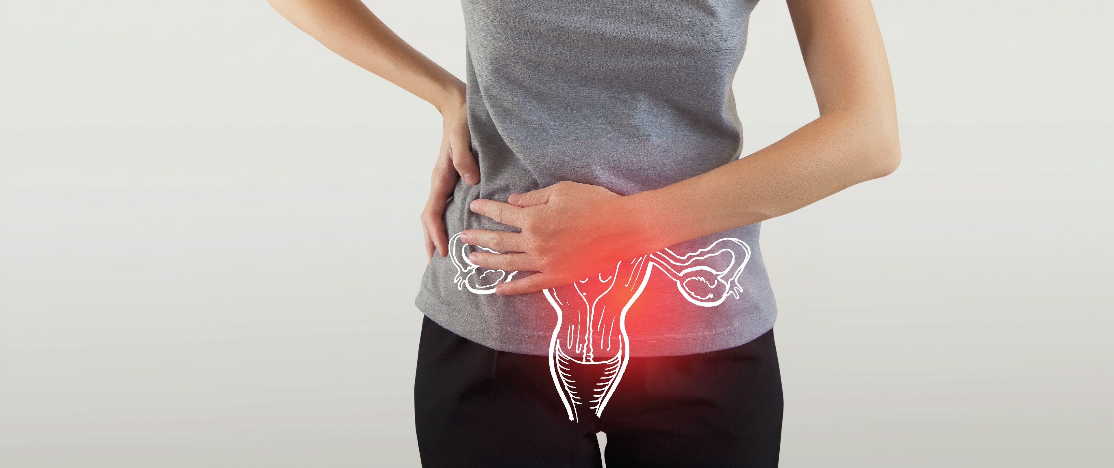
Uterine Fibroid Embolisation ( Ufe )
Promising minimally invasive treatment for uterine fibroids. The main indications are fibroids causing menorrhagia ( heavy bleeding ) or dysmenorrhoea ( severe pain during menses ) and adenomyosis. It is also indicated in cases of bleeding from the uterus due to uterine arteriovenous malformations or postpartum haemorrhage ( bleeding just after childbirth ) .
It is a minimally invasive day care procedure done under local anaesthesia through a small incision ( 2 – 3 mm ) in the groin area and involves stopping the blood supply to the fibroids.
The procedure is contraindicated in cases of malignancy, acute or chronic uterine infection and if the patient is pregnant.
- 85-90% of women who have had UFE experience significant or total relief of heavy bleeding, pain &/or bulk-related symptoms.
- All the fibroids are treated in one sitting
- UFE is effective for multiple & large fibroids. Recurrence of treated fibroids is very rare.
- Short and mid-term data show UFE to be very effective with a very low rate of recurrence.
- Can be done in patients unfit or unwilling for General or Regional anaesthesia
- Can be done as a daycare procedure / overnight stay
- Less postoperative complications
- Faster return to normal work
Embolisation Of Gastrointestinal Bleed
Bleeding from the stomach, small or large intestines is a major source of morbidity and sometimes mortality. In cases where bleeding does not stop by conventional means, abdominal angiography and embolisation provide a means of arresting the bleeding.
Abdominal angiography is a minimally invasive method to arrest bleeding in the stomach or intestines. Abdominal angiography is now recommended to not only indicate the site of bleed but to arrest the bleeding by closing the vessel ( embolisation ) that is supplying the area of bleeding. Therefore, it may avoid extensive surgery requiring removal of intestines.
Even in cases where eventually surgery is required, it is helpful in reducing morbidity by giving the surgeons an exact location of the diseased part of the bowel, thus avoiding unnecessary resection and reducing the time of surgery and postoperative complications.
Renal Interventions
- Percutaneous Nephrostomy / Antegrade Dj Stenting
Is indicated in patients with obstruction to urinary flow from the kidneys.
It involves puncturing the kidney directly to establish an alternative route for the flow of urine. It is especially indicated when DJ stenting is not possible / indicated. It is a daycare procedure and can also be done on an outpatient basis.
- Renal Angiography / Embolisation
Renal angiography and embolisation are indicated in patients with bleeding from the kidneys which is most commonly due to an intervention eg biopsy/surgery.
The bleeding vessel can be closed by embolisation techniques thus saving the patient another extensive surgery or in some cases removal of a kidney.
Embolisation of selective renal vessels can also be done for treatment of non – malignant conditions like angiomyolipomas or as a preoperative procedure in nephron-sparing surgery. It is a daycare procedure and same day discharge is possible.
- Renal Angioplasty/ Stenting
Is indicated when there is a narrowing of the renal artery causing the problem in renal function or high BP which is not controlled by medicines.
It involves opening the obstruction in the renal artery by a balloon dilatation ( angioplasty ) or in high-grade obstruction to putting a “scaffold” in the vessel ( stent ) to keep it open.
Graft Surveillance And Hemodialysis Access
It is crucial to extend the functional life of each access for as long as possible because the sites available for dialysis are limited and patients are dependant on dialysis for survival. Periodic surveillance venography can help increase the longevity of the graft/fistula
The incidence of renal diseases is increasing with a lot of patients requiring dialysis on a long term basis. The ideal hemodialysis access is an endogenous AV fistula or graft creation.
Graft surveillance is an established concept by the National kidney foundation – Dialysis Outcome Quality Initiative ( NKF – DOQI ) and involves regular assessment of flow through the graft. If flows through the graft are less than 400 – 600 ml/min, venography of the graft is recommended to study the inflow and outflow through the graft. Any obstruction to inflow or outflow can be corrected by interventional radiology techniques like balloon dilatation ( angioplasty ) or stenting of the narrowed portion of the vessel. These techniques can also help in the maturation of the fistula/graft.
In many cases, the fistula or graft does not mature or function optimally despite the best of efforts or there is an issue of multiple failed grafts. In these cases, a semi-permanent solution can be the insertion of a tunnelled hemodialysis catheter through which dialysis can be done for many months, till a more permanent graft or fistula creation can be made and it matures ( which requires on an average 6 weeks ). All these procedures are daycare procedures where same-day discharge is usually possible.
Arteriovenous Malformations ( AVM )
Endovascular or percutaneous therapy can be used to treat these subsets of diseases to achieve better cosmetic results/voiding surgery in larger lesions.
A wide range of techniques is available for treatment of this large group of lesions. Larger or deep-seated lesions will typically require endovascular embolisation therapy in which the vessel supplying the AVM is sealed off.
Smaller more superficial lesions can be adequately treated with percutaneous ablative therapies like alcohol and foam injections.
These techniques are less invasive with fewer complications than surgery. They also give a better cosmetic result. Typically they are OPD / daycare procedures with a one day stay required for extensive endovascular procedures.
Interventions In Liver Disorders
- Percutaneous Transhepatic Biliary Drainage ( Ptbd )
It is done in cases of obstruction to the outflow of bile in the liver especially where ERCP stenting is not possible, To relieve jaundice, an alternative channel can be created through a direct skin puncture and placement of a drainage tube in the biliary system.
Plastic and metallic stents can also be placed through the obstruction to keep the outflow tract open to allow free drainage of bile
It is a safe and effective day care procedure with same day discharge
- Transjugular Liver Biopsy ( TLB )
This is indicated when liver biopsy is essential but is not possible through direct puncture from the skin due to uncorrectable blood thinning, massive ascites ( fluid in the abdomen ), morbid obesity or grossly shrunken liver.
It involves taking a biopsy of the liver from within the body. a tiny puncture is made in a vein in the neck and through that vein, the liver is accessed and the biopsy was taken. It is an effective procedure ( diagnostic accuracy of approx 90 % ) with a far lesser complication rate in these subsets of patients than a direct percutaneous liver biopsy. It requires at the most an overnight stay in the hospital.
- Transjugular Intrahepatic Portosystemic Shunt ( Tips )
It is a procedure to establish an alternative channel for blood flow within the liver.
TIPS is most commonly performed for prevention of recurrent gastroesophageal variceal haemorrhage or refractory ascites in patients with severe portal hypertension, which cannot be managed successfully through endoscopic methods. It has also been used as a “ bridge to liver transplant".
Interventions In Cancer Patients
Interventional radiology techniques typically represent the least invasive but definitive diagnostic or therapeutic options available for patients with cancer. Many interventional radiology procedures can be performed on an outpatient basis or during a short hospital stay. Consequently, these procedures tend to be less expensive than other forms of therapy and frequently are associated with less risk and procedure-related complications, while giving equivalent results.
- Radio – Frequency Ablation ( Rfa )
RFA is a nonsurgical, localized treatment that kills the tumour cells with heat while sparing the adjacent healthy tissue.
In this procedure, the interventional radiologist guides a small needle through the skin into the tumour. From the generator, radiofrequency energy is transmitted to the tip of the needle, where it produces heat in the tissues and destroys it. The dead tumour tissue shrinks and slowly forms a scar. Depending on the size of the tumour, RFA can shrink or kill the tumour, extending the patient's survival time and greatly improving their quality of life while living with cancer.
The placement of the probe is guided by CT / ultrasound so that precise localisation is achieved and there is minimal damage to adjacent tissue. This reduces the chance of side effects and aids faster recovery. Thus, this treatment is much easier on the patient than conventional treatment methods and most people can resume their usual activities in a few days
RFA can also be used to treat benign conditions like osteoid osteoma ( type of a bone tumour) and it is very effective in alleviation of pain that is almost always associated with this condition. It is safe and effective and can spare the patient debilitating surgery. RFA is a day care procedure.
- Transarterial Chemo Embolisation ( Tace )
It is a useful therapy in the treatment of malignant liver lesions especially when the disease is widespread throughout the liver.
The procedure involves injecting chemotherapeutic agents mixed with small sponge particles into the artery that supplies the tumour. With this direct delivery technique, far lower dosages of the chemotherapeutic agent are needed than when the agent is delivered through the veins. This almost eliminates the side effects of chemotherapy. Concurrent injection of sponge particles cuts off the blood supply to a tumour which not only has an ischemic effect on the tumour itself but also prolongs the time that the chemotherapeutic agent is in contact with tumour cells, thus enhancing its efficacy. Patients who undergo transcatheter chemoembolization typically stay in the hospital for only one day
- Radio Embolisation ( Yttrium 90 Therapy )
Radioembolisation is the latest technique of intraarterial radiotherapy. It involves delivery of a strong dose of radiation therapy into the blood vessels supplying the tumour. It is indicated in extensive liver disease with bad liver function.
The procedure involves inserting a catheter in the blood vessels which supply a tumour and then injecting radioactive particles in these blood vessels. These microspheres get embedded in small blood vessels inside the tumour and emit beta particles ( radiation ) which treat a tumour.
Since the average penetration of a beta particle is only 2 mm, the radiation therapy delivered is highly targeted and is limited to the area of interest. This reduces the sometimes debilitating side effects of radiation therapy delivered in a conventional manner and increases the efficacy of the treatment as a far larger dose of radiation can be given to the tumour. It is useful even in large or multifocal liver tumours and in patients with substantially compromised liver function in whom other treatment modalities, even transarterial chemoembolisation is not an option. The procedure is done in two stages requiring one-day admission each.

Written and Verified by:

Related Blogs

Dr. Vivek Saxena In Radiology , Interventional Radiology
Nov 07 , 2020 | 1 min read

Dr. Vivek Saxena In Radiology , Interventional Radiology
Nov 07 , 2020 | 2 min read

Medical Expert Team
Nov 07 , 2020 | 1 min read
Blogs by Doctor
Hemoptysis: Know the underlying causes of coughing up blood
Dr. Vivek Saxena In Radiology , Interventional Radiology
Nov 07 , 2020 | 1 min read
Know about the minimally invasive treatment of fibroids!
Dr. Vivek Saxena In Radiology , Interventional Radiology
Nov 07 , 2020 | 2 min read
Most read Blogs


Related Blogs
Dr. Vivek Saxena In Radiology , Interventional Radiology
Nov 07 , 2020 | 1 min read
Dr. Vivek Saxena In Radiology , Interventional Radiology
Nov 07 , 2020 | 2 min read
Medical Expert Team
Nov 07 , 2020 | 1 min read
Blogs by Doctor
Hemoptysis: Know the underlying causes of coughing up blood
Dr. Vivek Saxena In Radiology , Interventional Radiology
Nov 07 , 2020 | 1 min read
Know about the minimally invasive treatment of fibroids!
Dr. Vivek Saxena In Radiology , Interventional Radiology
Nov 07 , 2020 | 2 min read
Most read Blogs